


Two grants totaling $3.7 million were awarded to San Diego State psychology professor Ralph-Axel Mueller from the National Institute of Mental Health.
These grants will assist Mueller in his ongoing research—working with brain imaging technology to detect neurological indicators of Autism Spectrum Disorder.
Presentation of the grants from the NIMH followed a rigorous application process.
“Very few grants are awarded without applying,” Mueller said. “With funding down in the past eight to 10 years, it is necessary to apply numerous times and be within the tenth percentile of your program to be considered.”
Since 2001, when Mueller began working for SDSU, he has strived to improve the process of diagnosing ASD and to discover by way of brain imaging that the neurological factors of the disorder that are present.
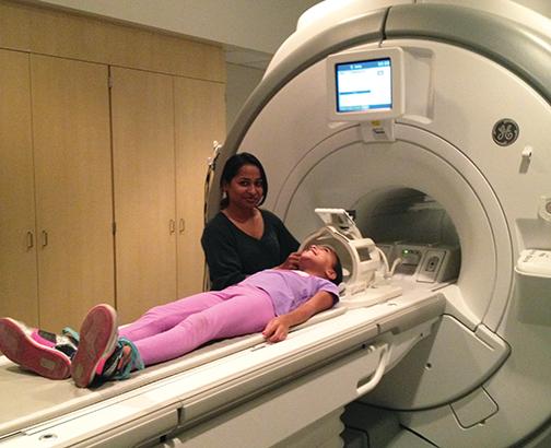
“Up until now, we’ve been using mostly MRI techniques, but there are some limitations with the temporal resolution,” Mueller said. “With MRI, you cannot get the big picture of what dynamically goes on in the brain but EEG and MEG are very good at that.”
Ultimately, Mueller hopes to uncover what is present in the brain to improve the process and accuracy of diagnosing ASD. Also, research will uncover which genetic risk factors are responsible.
“We are exploring what is wrong with brain networks in autism,” Mueller said. “It is currently diagnosed based on behavioral observation, but there is evidence that autism is a neurological disorder. Genes play a big role in autism but nobody really knows how at this point.”
Mueller will combine three brain-imaging technologies in his research with the money from the grant and will be working alongside two colleagues; Ksenija Marinkovic of SDSU and Tom Liu of University of California, San Diego.
Functional magnetic resonance imaging, magnetoencephalography and electroencephalography, are the specialized techniques of brain imaging that will be used together in the research.
“Our multimodal imaging approach is innovative as it benefits from the complementary advantages of the MEG/EEG and functional and structural MRI methods,” Marinkovic said. “It will provide multidimensional insight into functional, dynamic, and structural aspects of network connectivity in ASD.”
Each of the three imaging methods specialize in certain areas of the research.
“The MEG can provide more precise, spatial estimates of the underlying neural generators especially when combined with MRI images,” she said. “As a result, it can be used to examine neural communications in real time and at the level of an interactive system.”
MEG and EEG specialize with temporal resolution and explaining the temporal sequence with neural components.
“They are recorded simultaneously and reflect electrical potentials or magnetic fields associated with neural currents,” Marinkovic said.
Functional MRI specializes with research involving spatial resolution and vulnerable areas of individuals.
“It is also used to examine spontaneous interregional cross-correlations of the Blood Oxygenation Level Dependent signal that underlies fMRI,” she said.
The $3.7 million will go toward combining the three imaging methods for future strides in the field of research for ASD.
“A rich set of complementary measures including structural and functional MRI, anatomically-constrained MEG, and a comprehensive neuropsychological and behavioral battery will allow us to characterize the functional significance of expected network abnormalities with the aim of defining specific biomarkers for the ASD,” Marinkovic said.
Functional MRI specializes with research involving spatial resolution and vulnerable areas of individuals.
“It is also used to examine spontaneous interregional cross-correlations of the Blood Oxygenation Level Dependent (BOLD) signal that underlies fMRI,” she said.
The $3.7 million will go toward combining the three imaging methods for future strides in the field of research for ASD.
“A rich set of complementary measures including structural and functional MRI, anatomically-constrained MEG, and a comprehensive neuropsychological and behavioral battery will allow us to characterize the functional significance of expected network abnormalities with the aim of defining specific biomarkers for the ASD,” Marinkovic said.